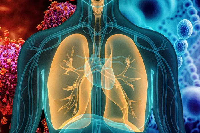
Bacterial pneumonia remains a serious respiratory infection requiring accurate diagnosis and prompt treatment. Distinguishing bacterial pneumonia from viral cases and other lung conditions ensures patients receive appropriate care. Medical professionals use a combination of clinical evaluation, imaging studies, and laboratory tests to confirm bacterial lung infections.
Recognizing Common Symptoms
Bacterial pneumonia typically presents with distinct symptoms that develop over hours to days. A persistent cough producing yellow, green, or bloody mucus often indicates bacterial involvement. The sputum tends to be thicker and more colored than with viral infections.
Fever frequently spikes above 101°F (38.3°C) and may be accompanied by chills and sweating. Patients often describe sharp chest pain that worsens with deep breathing or coughing. Shortness of breath develops as lung tissue becomes inflamed and less functional.
Physical Examination Findings
Doctors listen carefully to the lungs through a stethoscope. Bacterial pneumonia typically causes crackling or bubbling sounds called rales as air moves through fluid-filled alveoli. Dullness to percussion over the affected area suggests lung consolidation.
Increased respiratory rate above normal levels indicates the body’s struggle to oxygenate blood. Bluish lips or nail beds signal oxygen deprivation requiring urgent attention. Mental confusion in elderly patients may be the primary symptom rather than typical respiratory complaints.
Chest Imaging Techniques
Chest X-rays remain the primary imaging tool for pneumonia diagnosis. Bacterial pneumonia often shows lobar consolidation – a distinct white area affecting one lung section. Air bronchograms may appear as dark branching lines within the consolidated area.
CT scans provide more detailed images when X-rays prove inconclusive or complications are suspected. These scans better visualize lung abscesses or pleural effusions. Imaging also helps assess treatment response by showing resolution of infiltrates.
Laboratory Blood Tests
Complete blood counts typically show elevated white blood cells with increased neutrophils in bacterial pneumonia. Very high or very low counts may indicate severe infection. C-reactive protein and procalcitonin levels help distinguish bacterial from viral causes.
Blood cultures identify bacteria spreading into the bloodstream, though they’re positive in only 20-30% of cases. Arterial blood gases measure oxygen and carbon dioxide levels, important for assessing respiratory function in severe cases.
Sputum Analysis
Examining coughed-up mucus can reveal causative bacteria. Gram staining provides rapid results about bacterial type – gram-positive or gram-negative. Culture and sensitivity testing identifies specific organisms and determines effective antibiotics.
Proper sputum collection is crucial, as saliva samples won’t show lung bacteria. Deep cough specimens collected before antibiotics yield the best results. Some bacteria like Streptococcus pneumoniae may be part of normal throat flora, requiring careful interpretation.
Urinary Antigen Tests
Rapid urine tests detect specific bacterial components from Streptococcus pneumoniae and Legionella pneumophila. These provide quick results even after antibiotic initiation. The tests are particularly useful for severe cases requiring immediate treatment guidance.
Pneumococcal urinary antigen tests remain positive for weeks after infection, helping diagnose cases with prior antibiotic use. Legionella urine antigen testing only detects serogroup 1, the most common disease-causing strain.
Bronchoscopy Procedures
When standard tests don’t identify the cause or treatment fails, bronchoscopy may be performed. This procedure uses a thin tube with a camera to examine airways and collect samples. Protected specimen brushing and bronchoalveolar lavage obtain uncontaminated lower respiratory samples.
Bronchoscopy helps diagnose unusual pathogens in immunocompromised patients. It also evaluates for alternative diagnoses like tumors or foreign bodies. The procedure carries more risk than other diagnostic methods and requires specialized equipment.
Pleural Fluid Analysis
When pneumonia causes fluid accumulation around the lung (pleural effusion), analyzing this fluid can aid diagnosis. Thoracentesis removes fluid for testing using a needle. Bacterial pneumonia typically causes exudative effusions with high protein and white blood cell counts.
Pleural fluid pH, glucose, and lactate dehydrogenase levels help determine infection severity. Gram stain and culture of pleural fluid may identify organisms when blood and sputum tests fail. This procedure also provides therapeutic relief for large effusions.
Severity Assessment Tools
Doctors use scoring systems to determine pneumonia severity and guide treatment decisions. The CURB-65 score evaluates confusion, urea levels, respiratory rate, blood pressure, and age. Higher scores indicate greater mortality risk and need for hospitalization.
The Pneumonia Severity Index (PSI) incorporates more variables to predict complications. These tools help avoid unnecessary hospitalizations for low-risk patients while ensuring sicker patients receive appropriate care.
Differential Diagnosis Considerations
Several conditions mimic bacterial pneumonia and must be ruled out. Viral pneumonia typically causes less severe symptoms and shows different patterns on imaging. Pulmonary edema from heart failure may cause similar breathlessness but with different exam findings.
Pulmonary embolisms can produce chest pain and hypoxia without infection signs. Lung cancer sometimes appears as persistent infiltrates on imaging. Tuberculosis should be considered in chronic cases or high-risk populations.
Special Patient Populations
Diagnostic approaches vary for different groups. Elderly patients may show atypical presentations with minimal fever and more confusion. Young children often have higher fevers but may not produce sputum for testing.
Immunocompromised patients risk infections from unusual organisms requiring specialized tests. Hospitalized patients may develop pneumonia from different bacteria than community-acquired cases. These variations influence testing and treatment choices.
When to Suspect Treatment Failure
Lack of improvement after 48-72 hours of antibiotics suggests possible diagnostic error. Persistent fever, worsening symptoms, or new complications indicate need for reevaluation. Imaging may reveal abscesses, empyema, or expanding infiltrates requiring treatment changes.
Secondary infections or incorrect initial diagnosis sometimes explain treatment failures. Repeat testing including cultures and imaging helps identify the cause. Some bacteria resist standard antibiotics, requiring alternative regimens.
Emerging Diagnostic Technologies
New methods are improving pneumonia diagnosis. Molecular tests like PCR rapidly identify pathogens from respiratory samples. Mass spectrometry speeds up organism identification from cultures. Biomarker panels help distinguish bacterial from viral causes.
Point-of-care ultrasound allows bedside lung evaluation, especially useful in critical care. Artificial intelligence assists in interpreting chest imaging. These advances promise faster, more accurate diagnoses in coming years.
Importance of Timely Diagnosis
Early accurate diagnosis improves bacterial pneumonia outcomes. Appropriate antibiotic selection reduces complications and hospitalization duration. Delayed treatment increases risks of sepsis, respiratory failure, and death.
Proper diagnosis also prevents unnecessary antibiotic use for viral cases, combating resistance. Identifying specific pathogens guides infection control measures in healthcare settings. Follow-up testing confirms resolution and detects lingering abnormalities.
Conclusion
Diagnosing bacterial pneumonia requires synthesizing clinical findings, test results, and patient factors. No single test provides perfect accuracy, making clinical judgment essential. Understanding diagnostic methods helps healthcare providers deliver targeted, effective care.
Ongoing research continues refining diagnostic tools and approaches. Combining traditional methods with emerging technologies enhances our ability to identify and treat bacterial pneumonia promptly. Accurate diagnosis remains the critical first step toward successful treatment and recovery.
Related topics:
How To Get Rid Of Bacterial Infection In Nose?
Treating Bacterial Infections: A Comprehensive Guide
What Can You Give an Infant for a Cough?


